
 Dental Imaging
Dental Imaging
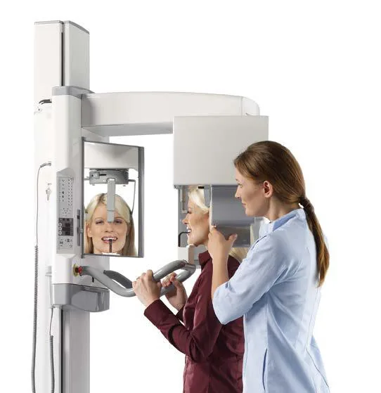
Soredex CRANEX D Digital Panoramic and Cephalometric X-ray Unit
- Model
- CRANEX D
- Brand
- Soredex
Soredex CRANEX D digital panoramic and cephalometric system with a fast 11-second scan, 50% thicker anterior layer and left or right Ceph option.
Key features
- Fast 11-second panoramic scan and HiQ scan of about 17 seconds
- Soredex-patented Variable Panoramic Collimator (VPC) with an anterior layer 50% thicker than conventional methods
- Automatic Exposure Setting (AES) suggesting exposure values based on the patient's head size
- Automatic spine shadow compensation in all panoramic programs
- 5-point head support, self-locking temple supports and three positioning lasers
- Open design for wheelchair access and imaging in standing and seated positions
- Cephalometric geometry with true central projection and constant magnification without distortion
- Left-hand or right-hand Ceph option, with single-sensor or dual-sensor design
Description
CRANEX D is a modern digital panoramic and cephalometric imaging system built for today's high-volume, fast-paced dentistry. Clear, accurate digital images together with stable, comfortable patient positioning make everyday diagnosis simple and precise.
Fast panoramic programs suit follow-up patients and busy clinics, while High Quality (HiQ) programs are designed for more demanding diagnostic cases. Cephalometric capability can be added on site at any time.
Technical specifications
| Cephalometric field | Full width: 22 x 26 cm, Reduced width: 22 x 18 cm |
|---|
| Sensor | CCD |
|---|---|
| Sensor active area | PAN: 147.5 x 6.1 mm, CEPH: 221.2 x 6.1 mm |
| Sensor / image pixel size | 48 µm / 96 µm |
| File size | PAN max 9.5 MB, CEPH max 11.5 MB |
|---|
| Generator | High frequency DC, operating frequency 40 kHz |
|---|---|
| Tube voltage | 57 to 85 kV |
| Tube current | 10 mA |
| Exposure time | 17.6 s panoramic HiQ, 11 s fast panoramic, 8 to 20 s cephalometric |
| Focal spot | 0.5 mm |
|---|---|
| Minimum total filtration | 2.7 mm Al |
| SID | 520 mm panoramic, 1721 mm cephalometric |
| Input voltage | 230/240 Vac ±10% / 115 Vac (50/60 Hz) |
|---|---|
| Fuse | 8 A / 16 A slow (230/115 Vac) |
| Electrical safety class | EN 60601-1 class 1/B |
| Weight | Pan 120 kg, Ceph 165 kg |
|---|
Applications
- Adult and pediatric panoramic (pediatric with shorter exposure time and smaller field)
- Extraoral bitewing, bilateral or unilateral
- Sectional imaging with selection of 1 to 5 segments for dose reduction
- TMJ and sinus
- Lateral and PA cephalometry
- Carpus (optional, with CRANEX D Ceph)
Accessories
- Cephalometric module upgradeable on site (left or right)
- DIGORA for Windows with optional network version and DICOM
- SCANORA SW with network license and DICOM
- SOREDEX TWAIN and SorCom (DICOM bridge)
- CliniView XV
Downloads
Related products
- Dental Imaging
 Dental Imaging
MoritaVeraviewepocs 2D
Morita Veraviewepocs 2D Digital Panoramic and Cephalometric Unit
Details Inquiry Dental Imaging
 Dental Imaging
Related knowledge
View all
Tech tip · Dental Imaging
Planmeca ProMax Cephalostat with ProTouch User's Manual
User's manual for the Planmeca ProMax Cephalostat with ProTouch touchscreen, for accurate head positioning and cephalometric imaging on 2D and 3D ProMax units.
Planmeca
Tech tip · Dental Imaging
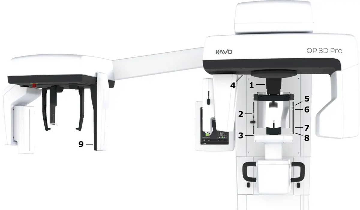
ORTHOPANTOMOGRAPH OP 3D Pro User Manual
User manual and technical specifications for the OP 3D Pro digital dental imaging system with panoramic, cephalometric and Cone Beam 3D programs.
Instrumentarium Dental