

 Dental Imaging
Dental Imaging
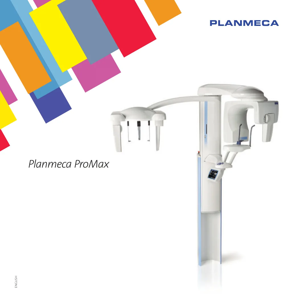
Planmeca ProMax Panoramic, Cephalometric and Tomographic X-ray Unit
- Model
- ProMax
- Brand
- Planmeca
Planmeca ProMax extraoral imaging with SCARA robotics for panoramic, TMJ, sinus, 2D tomography and cephalometry, upgradeable to 3D.
Key features
- SCARA robotic technology with real-time calculation of rotation patterns for each patient
- Two models: SCARA3 (all programs including tomography and 3D) and SCARA2 (panoramic, TMJ and sinus, upgradeable with a third joint)
- Three laser beams (midsagittal, Frankfort and focal layer) with no need for a mirror
- Dynamic Exposure Control (DEC) in digital panoramic and cephalometric imaging
- Autofocus with a short scout exposure to determine the focal layer automatically
- Adjustable focal layer shape based on the size and shape of the patient's jaw
- Automatic removal of the cervical spine shadow in the anterior region
- Child mode with 35% dose reduction, and horizontal and vertical segmenting with dose reduction up to 93%
Description
Planmeca ProMax offers a wide range of extraoral imaging modalities: panoramic imaging of the dental arch, maxillary sinus, temporomandibular joint, 2D linear tomography and cephalometry. With a three-axis SCARA robotic arm, the platform delivers the highly precise movements required in rotational maxillofacial radiography without mechanical limitations.
The open, side-entry positioning concept allows easy access for all patients, including wheelchair patients, and direct eye contact with the operator. Every Planmeca ProMax, film or digital, can be upgraded to Planmeca ProMax 3D s or ProMax 3D by changing the sensor and updating the software.
Technical specifications
| CCD pixel size | 33 µm |
|---|---|
| Image pixel size | 66 / 99 / 132 µm selectable |
| CCD active area | Pan 9 x 136 mm, Ceph 9 x 270 mm |
| Magnification | Pan constant 1.2, Ceph 1.08 to 1.13 |
|---|---|
| Digital resolution | Pan max 9 lp/mm, Ceph max 5.7 lp/mm |
| Digital image field | Pan 14 x 30 cm, Ceph 24/27 x 18/30 cm |
| File size | Pan 4.5 to 7.7 MB, Ceph 5 to 12 MB |
| Generator | Constant potential, resonance mode high frequency 80 to 150 kHz |
|---|---|
| Tube voltage | 50 to 84 kV |
| Tube current | 0.5 to 16 mA DC |
| Exposure time | Pan 2.7 to 16 s, Ceph 0.2 to 19 s, Tomo 3 to 24 s per frame |
| X-ray tube | D-054SB-P |
|---|---|
| Focal spot | 0.5 x 0.5 mm (IEC 336) |
| Filtration | Minimum 2.5 mm Al |
| SID | Pan 500 mm, Ceph 163 to 170 cm |
| Input voltage | 100 to 240 V, 50 or 60 Hz |
|---|
| Panoramic dimensions | 96 x 125 x 153-243 cm (W x D x H) |
|---|---|
| Dimensions with cephalostat | 194 x 125 x 153-243 cm (W x D x H) |
| Weight | 113 kg, with cephalostat 128 kg |
Applications
- Standard panoramic, Interproximal (caries detection), Orthogonal (periodontal and implant) and Bitewing
- TMJ lateral and PA, Double TMJ and Multi-angle TMJ
- Maxillary sinus imaging
- Cross-sectional and longitudinal tomography for implants, wisdom teeth and TMJ
- Lateral, PA and hand cephalometry with a field up to 30 x 27 cm
Accessories
- Digital cephalostat with shared or dedicated sensor
- Planmeca ProMax Cephalostat CA (automatic) or CM (manual) for the film version
- Accessory cabinet
- External graphical interface
- Planmeca Romexis software for Windows and Mac OS with full DICOM 3.0 support
- Upgrade to Planmeca ProMax 3D s or ProMax 3D, with a 3D face scan option
Downloads
Related products
- Dental Imaging
 Dental Imaging
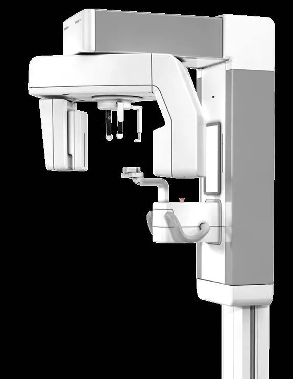
MoritaVeraviewepocs 2D
Morita Veraviewepocs 2D Digital Panoramic and Cephalometric Unit
Details Inquiry Dental Imaging
 Dental Imaging
Related knowledge
View all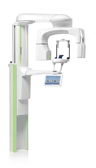
Tech tip · Dental Imaging
Planmeca ProMax Cephalostat with ProTouch User's Manual
User's manual for the Planmeca ProMax Cephalostat with ProTouch touchscreen, for accurate head positioning and cephalometric imaging on 2D and 3D ProMax units.
Planmeca
Tech tip · Dental Imaging
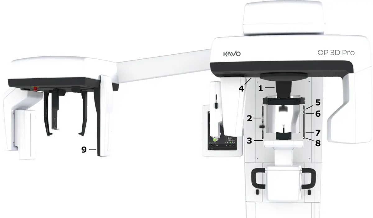
ORTHOPANTOMOGRAPH OP 3D Pro User Manual
User manual and technical specifications for the OP 3D Pro digital dental imaging system with panoramic, cephalometric and Cone Beam 3D programs.
Instrumentarium Dental