

 Dental Imaging
Dental Imaging
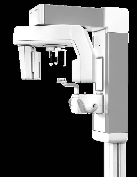
Morita Veraviewepocs 2D Digital Panoramic and Cephalometric Unit
- Model
- Veraviewepocs 2D
- Brand
- Morita
High-speed Veraviewepocs 2D digital panoramic and cephalometric unit with 7.4 s scan time, AF auto positioning and upgradeability to Veraviewepocs 3D.
Key features
- Fine High Speed panoramic with 7.4 s exposure time and about 1/4 the radiation compared with the Veraviewepocs film system
- High-speed cephalometry in about 4.9 s with about 1/10 the radiation, completing the imaging process in 20 seconds
- Digital Direct Automatic Exposure (DDAE) controls kV and mA simultaneously and improves dynamic range
- Automatic Image Enhancer (AIE) reveals detail in very bright or dark areas
- AF auto positioning with PSD sensor and 0.2 mm accuracy for high reproducibility
- Three laser beams (Frankfurt plane, sagittal plane and image layer) for precise positioning
- Motorized C-arm movement with soft start and stop and automatic stop on overload
- Carbon temple rods with minimal shadow in the image
Description
Veraviewepocs 2D is a high-speed digital panoramic and cephalometric unit that delivers very high quality images at a lower radiation dose. Panoramic exposure time is 7.4 seconds and cephalometric exposure time is about 4.9 seconds.
Specialized programs such as Orthoradial Panoramic to reduce overlapping of adjacent teeth and Shadow Reduction Panoramic to reduce interfering shadows, together with AF auto positioning, make accurate diagnosis easier.
The unit is fully upgradeable to the Veraviewepocs 3D R100 / F40.
Technical specifications
| Model | X550, 2D (panoramic) and 2D CP (panoramic / cephalometric) versions |
|---|---|
| Positioning | Electric motor and AF optical distance sensor with 0.2 mm accuracy |
| Cephalometric scan time | Approx. 4.9 s |
| Panoramic sensor | CCD with 32-bit processor |
|---|---|
| Pixel size | 144 µm (Fine High Speed), 96 µm (Super Fine) |
| Cephalometric sensor | CCD, 225 mm height, digital cassette shared between panoramic and cephalometric |
| Panoramic magnification | 1.3 / 1.5 / 1.6 |
|---|---|
| Cephalometric image area | LA 225 x 254 mm, PA 225 x 203 mm |
| Cephalometric magnification | 1.1 |
| Tube voltage | 60 to 90 kV (DDAE or manual) |
|---|---|
| Tube current | 1 to 10 mA (DDAE or manual) |
| Panoramic exposure time | Approx. 7.4 s (Fine High Speed), approx. 15 s (Super Fine) |
| Effective focal spot | 0.5 mm |
|---|
| Power supply | 220 / 230 / 240 V, 50/60 Hz |
|---|---|
| Power consumption | 2.3 kVA |
| Dimensions | 2D: 1020 x 1330 x 2355 mm, 2D CP: 2000 x 1330 x 2355 mm (W x D x H) |
|---|---|
| Installation space | 2D: 1.35 m2, 2D CP: 2.60 m2 |
| Weight | 2D: approx. 190 kg, 2D CP: approx. 258 kg |
| Wheelchair access width | Up to 480 mm |
Applications
- Standard Panoramic with constant 1.3x and 1.6x magnification
- Pedodontic Panoramic for children and small jaws at lower dose
- Orthoradial Panoramic to reduce tooth overlap
- Shadow Reduction Panoramic to reduce the mandibular ramus shadow
- Maxillary Sinus Panoramic with 1.5x magnification
- TMJ 4 Views with the beam angle aligned to the condyle axis
- Digital Lateral and PA cephalometry
Accessories
- Optional wall-mounted version for wheelchairs wider than 480 mm
- Upgrade to Veraviewepocs 3D R100 / F40 with 3D FOVs from Ø 40 x 40 to Ø R100 x 80 mm
Downloads
Related products
- Dental Imaging
 Dental Imaging
 Dental Imaging
 Dental Imaging
Related knowledge
View all
Tech tip · Dental Imaging
Planmeca ProMax Cephalostat with ProTouch User's Manual
User's manual for the Planmeca ProMax Cephalostat with ProTouch touchscreen, for accurate head positioning and cephalometric imaging on 2D and 3D ProMax units.
Planmeca
Tech tip · Dental Imaging
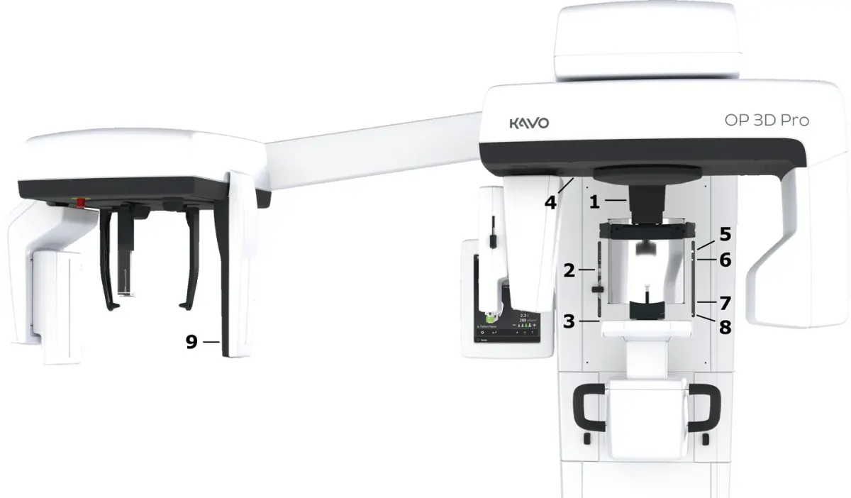
ORTHOPANTOMOGRAPH OP 3D Pro User Manual
User manual and technical specifications for the OP 3D Pro digital dental imaging system with panoramic, cephalometric and Cone Beam 3D programs.
Instrumentarium Dental