

 Imaging Software
Imaging Software
MUSICA™ image processing
- Model
- MUSICA image processing
- Brand
- Agfa
Your first image is the right one: MUSICA™ image processing with Fractional Multiscale Processing for chest, abdomen, extremities, spine, pelvis and neonatal exams.
Description
Your first image is the right one.
Why choose MUSICA™ for chest exams?
- Increased visualization of catheters, tubes and PICC lines with the catheter companion image
- Visible microfractures and nodules behind dense structures with digital tomosynthesis
- High detail in the mediastinum with MUSICA™ 3
Why choose MUSICA™ for abdomen exams?
- Sharp delineation of the bowel wall with MUSICA™ 3+
- Clear, simultaneous presentation of both dense and non-dense areas
- Improved visualization of abdominal organs and of catheters, stents, etc.
- Differentiation of skin and fat layers in the periphery
Why choose MUSICA™ for extremity exams?
- Sharp trabecular and cortical bone
- Balanced presentation of both soft tissue and overlapping bone structures
- True representation of implants with clear bone interfaces
Why choose MUSICA™ for spine and pelvis exams?
- Increased image quality for bariatric patients
- More detail in relevant skeletal structures
Why choose MUSICA™ for neonatal exams?
- Optimized visualization of organ boundaries and free air
- Clear lung markings
- Sharp bone outline
- High visibility of low-contrast structures like PICC lines
MUSICA™ also works in dynamic imaging
Dynamic MUSICA™ brings enhanced temporal noise suppression as well as brightness and density stabilization to dynamic imaging studies.
How does MUSICA™ 3 work?
MUSICA’s Fractional Multiscale Processing technology automatically analyzes the layers of each raw image and optimizes the processing parameters, independent of body part or dose deviation. It enhances noise suppression, offers brightness control, reduces veiling glare and plays a significant role in enabling potential dose reduction. The result is excellent image quality compared to standard image processing, for both bone and soft tissue.
To render the most difficult zones of an image, such as abrupt transitions from low- to high-density areas, a mathematical algorithm called Fractional Multiscale Processing (FMP) is applied. With this algorithm, the image processing filters are further decomposed into elementary fractions, which are processed separately. As a result, grayscale differences are represented in a more natural way, without artifacts.
FMP also eliminates the need for window/level adjustment to enhance the visibility of details.
“Subtle bone details often tend to fade in the vicinity of implant edges, but with the next-generation MUSICA, these details are well preserved and easily visible. I compare it to being able to hear a pianissimo passage after an explosion.”
What our customers say
“The image quality is exceptional. The fact that we don’t need to adjust the images has a lot of advantages: image quality, workflow, efficiency… we save a lot of time. And we are also getting a consistent appearance to our images, which is important for referrers and clinicians.”
“MUSICA image processing is so sharp, so much sharper than people are used to! You can identify things you just couldn’t see before.”
White papers
MUSICA® catheter processing enhances neonatal imaging and care
A study evaluating the ability of image processing to improve the visualization of central catheter tip positioning in newborns was published in BMC Medical Imaging. The authors found that Agfa’s MUSICA® Catheter Processing with Fractional Multiscale Processing (FMP) technology significantly improved the perception of low-contrast PICC line tips in digital chest X-rays, compared to standard image processing.
Bedside imaging without a grid
In a clinical evaluation, MUSICA3™+ non-grid images were rated significantly higher than MUSICA2™ images acquired with or without a grid.
For mobile non-grid imaging, optimal lung field visualization, as provided by MUSICA3™+, was seen by the radiologists as the most important aspect of a bedside chest examination. This potentially outweighs the additional improvement of the mediastinum gained by using a physical anti-scatter grid with MUSICA2™ general radiography processing at a 1.6x higher exposure.
Dose reduction with MUSICA™
A study involved five hospitals in Germany, six independent radiologists and data collected over more than two years. All relevant body parts and exam types, for both adult and pediatric X-ray imaging, were included.
The conclusion is that only half of the dose prescribed by Germany’s national diagnostic reference levels (NDRLs) is required for close-to-maximum image quality and diagnostic confidence. The study also confirms with high statistical confidence (95% confidence level, 80% power) the diagnostic usability and quality of images created by Agfa DR systems using MUSICA™ and CsI DR detector technology.
Digital tomosynthesis
Several musculoskeletal cases are presented with indications and imaging examples (2D and DTS). The main advantages of Agfa’s digital tomosynthesis can be summarized as follows:
- Improved triage, by making the X-ray exam more definitive
- Little or no patient repositioning is required to obtain the necessary clinical information
- A significantly lower dose is required compared to CT, even low-dose CT
External references
Notohamiprodjo, S., Roeper, K.M., Mueck, F.G. et al. Advances in multiscale image processing and its effects on image quality in skeletal radiography. Sci Rep 12, 4726 (2022). doi:10.1038/s41598-022-08699-8
Applications
- Chest exams
- Abdomen exams
- Extremity exams
- Spine and pelvis exams
- Neonatal exams
- Dynamic imaging
Related products
- Imaging Software
 Imaging Software
 Imaging Software
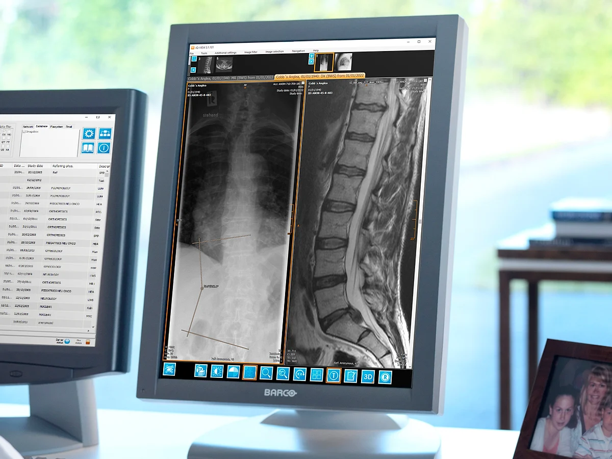 Imaging Software
Related knowledge
View all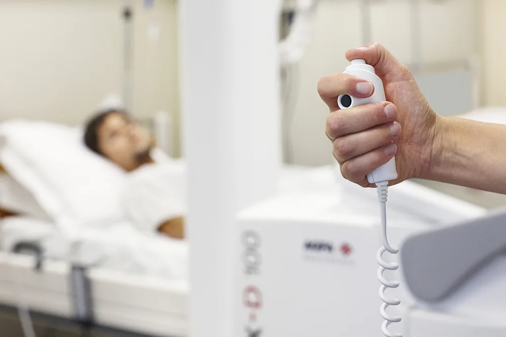
News · X-ray Systems
Transforming Radiography Services at NHS Lothian with Agfa’s DR 600 X-ray room
Modern imaging departments need technology that not only delivers exceptional image quality but also supports staff efficiency and patient care. At NHS Lothian’s Royal Infirmary of Edinburgh, the tran...
2 min readAgfa
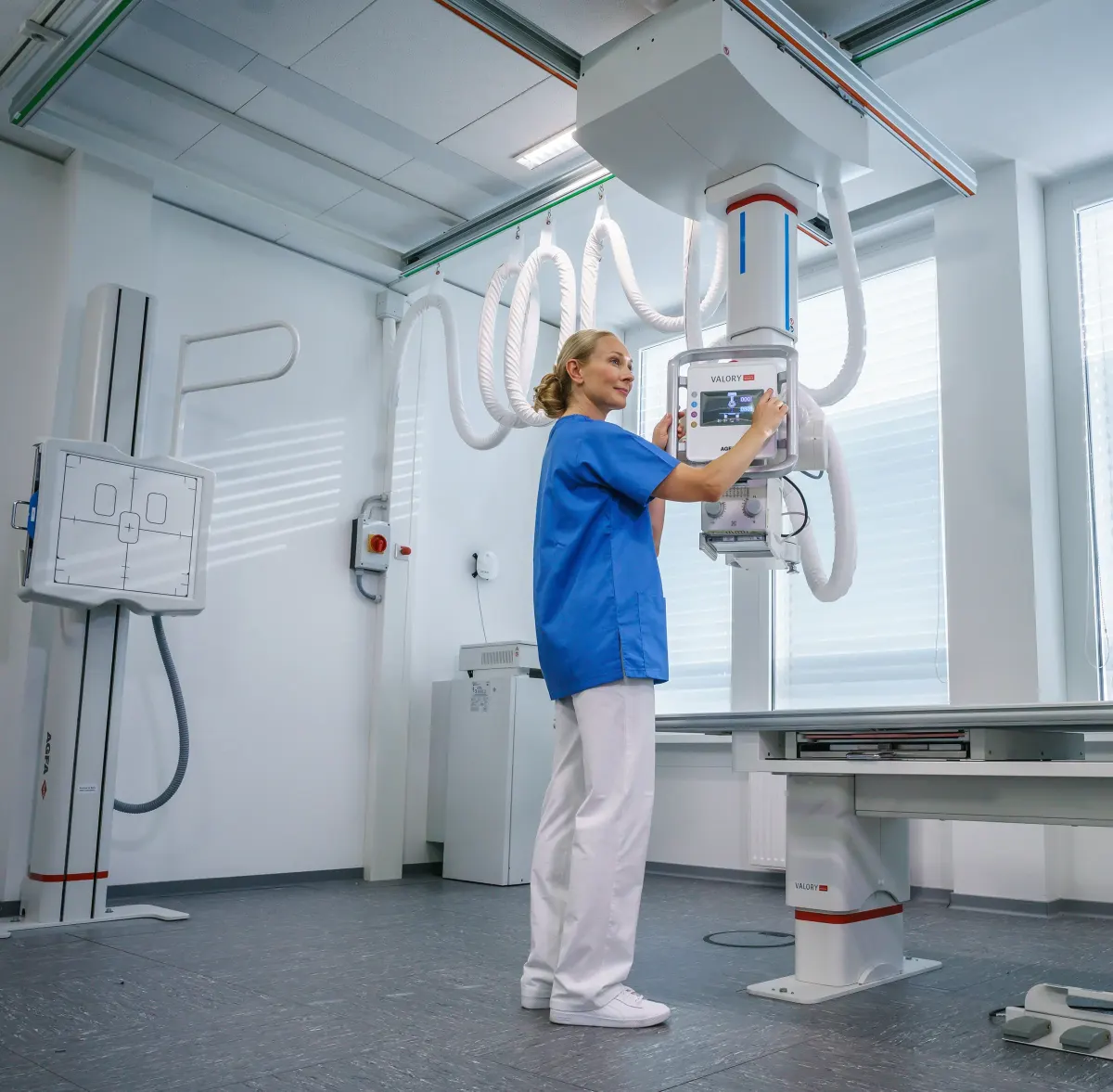
News · Portable X-ray
At Poland’s PLTR 2023 Congress, Agfa demonstrates how it “Empowers X-ray Experts” with meaningful answers to their daily needs
Agfa’s booth at PLTR 2023 offers visitors an opportunity to discover smart, meaningful answers to radiology’s real needs. In addition to solutions physically present at the booth, Agfa’s portfolio wil...
1 min readAgfa
News · Digital Radiography
Bumi Medika Ganesha Clinic ITB, West Java, installs Agfa’s DX-D 300 U-arm solution with Dura-line detector.
Early last month, the Bumi Medika Ganesha Clinic ITB (institut Teknologi Bandung), West Java, Indonesia installed Agfa’s DX-D 300 U-arm solution. Agfa’s DX-D 300 offers a fast way to go digital, easil...
1 min readAgfa