

 Mammography
Mammography
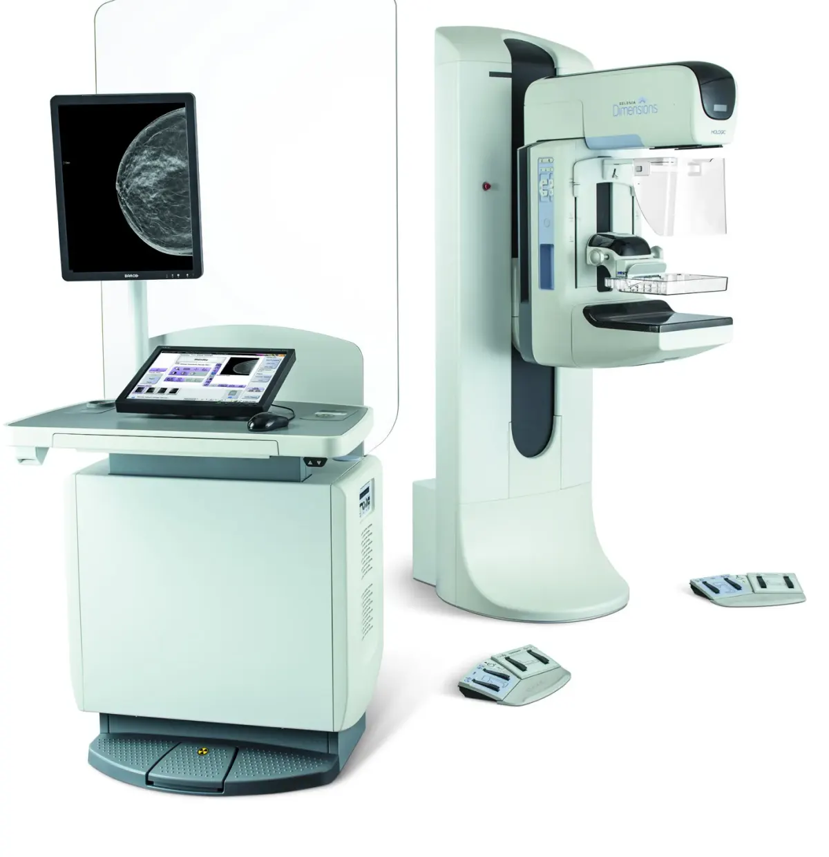
Hologic Selenia Dimensions Mammography and Tomosynthesis System
- Model
- Selenia Dimensions
- Brand
- Hologic
Hologic Selenia Dimensions digital mammography with Genius 3D MAMMOGRAPHY, 70 micron amorphous selenium detector, 3D biopsy and Contrast Enhanced 2D.
Key features
- Genius 3D MAMMOGRAPHY with Combo, TomoHD and ComboHD modes
- C-View to generate 2D images from 3D data and reduce dose
- Contrast Enhanced 2D (CE2D) with I-View software, alone or combined with 3D Diagnostic
- Stereotactic and tomosynthesis biopsy with the Affirm system
- Direct-capture amorphous selenium detector and HTC cellular grid that retracts for 3D and magnification views
- Split C-arm with vertical motion and motorized rotation
- Quantra breast density assessment and CAD with ImageChecker (2D, C-View and 3D)
- Ergonomic packages with motorized height adjustment, fingerprint login and barcode reader (depending on package)
Description
Selenia Dimensions is Hologic's digital mammography system, designed from the ground up for breast tomosynthesis, offering high image quality, high productivity and a range of advanced applications. The Genius 3D MAMMOGRAPHY exam is designed for earlier detection of breast cancer, clearer images of lesions, and fewer recalls and biopsies.
The platform is available in several packages (Avia 3000 for 2D only, 6000 and 9000) and can be upgraded as the facility's needs grow, from low-dose 3D imaging and 3D biopsy to Contrast Enhanced 2D co-registered with tomosynthesis.
Technical specifications
| 3D scan angle | 15° with 15 projection images |
|---|---|
| 3D scan time | 3.7 s |
| Cycle time | 26 to 42 s depending on mode |
| C-arm rotation | 2D: +195° to -155°, 3D and biopsy: +180° to -140° |
| Full compression force | 89 to 178 N |
| Monitor | 2 MP color or 3 MP medical grayscale |
| Detector | TFT-based direct capture, amorphous selenium |
|---|---|
| Detector size | 24 cm x 29 cm |
| Pixel size | 70 microns |
| Spatial resolution | 2D: 7.1 lp/mm, 3D: 3.5 lp/mm |
|---|---|
| Image bit depth | 14-bits |
| Magnification factors | 1.5x and 1.8x |
| Image buffer capacity | About 9,000 four-view 2D studies, about 3,000 3D studies |
| Generator | High Frequency Inverter, 7.0 kW, 200 mA @ 35 kV |
|---|---|
| Tube voltage | 2D: 20 to 39 kV, 3D: 20 to 49 kV |
| mAs range | 3.0 to 500 mAs |
| SID | 70 cm |
|---|---|
| X-ray tube | Bi-angular rotating tungsten anode, 9500 RPM, 222 kJ (300,000 HU) |
| Focal spot | 0.3 mm and 0.1 mm |
| Filtration | Rh 0.05 mm, Ag 0.05 mm, Al 0.70 mm (3D), Cu 0.30 mm (CE2D) |
| Storage | 8 GB (Avia 3000) or 16 GB (6000/9000) RAM, 1.0 TB |
|---|
| Connectivity | DICOM Print, Query, Storage, Storage Commitment, Worklist; IHE |
|---|
| Power supply | 200-240 VAC, single phase, 50/60 Hz |
|---|
Applications
- 2D and 3D screening mammography
- 2D and 3D diagnostic mammography
- Stereotactic and tomosynthesis biopsy
- Contrast Enhanced 2D imaging
- Mobile units (with mobile coach kit)
Accessories
- Screening compression paddles (24x29, 18x24, small breast)
- Contact, spot, magnification and localization paddles
- Ultrasound paddle
- Carbon fiber magnification platform and localization cross-hair
- Two dual-function footswitches
- Affirm biopsy system, Cenova server and Advanced Workflow Manager
Downloads
Related products
- Mammography
 Mammography
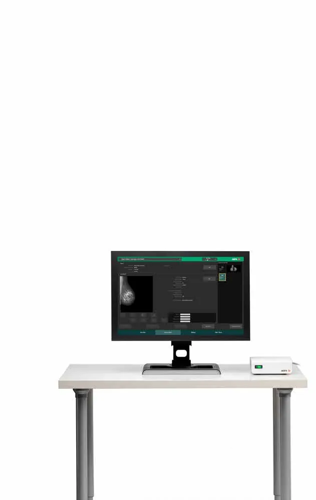
AgfaMammo DR Retrofit (DR 24M / DR 18M)
Agfa Mammo DR Retrofit Mammography Upgrade Solution
Details Inquiry Mammography
Related knowledge
View all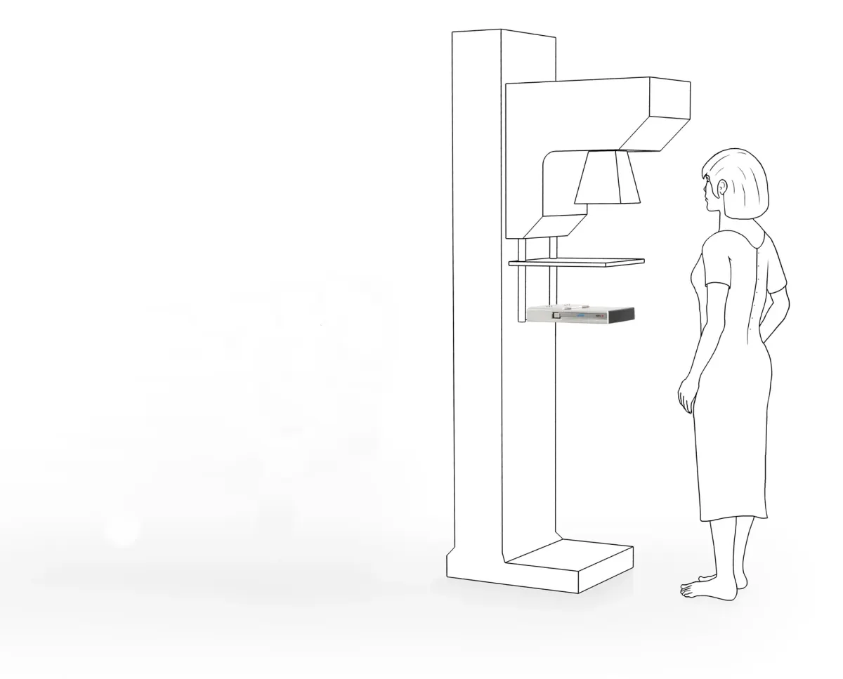
Article · Mammography
Agfa Mammography Portfolio
Agfa's vendor-neutral mammography portfolio: Mamoray film, DRYSTAR printers, MUSICA processing, CR Mammo and DR Mammo Retrofit.
Agfa
Article · Mammography
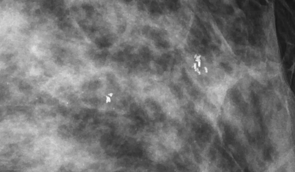
Does the Pixel Size of a Full-Field Digital Mammography Detector Matter for Early Detection of Breast Cancer?
Agfa white paper comparing FFDM, retrofit and CR systems with 50 to 100 µm pixels, showing pixel size alone does not determine detectability.
3 min readAgfa