

Article3 min read
Does the Pixel Size of a Full-Field Digital Mammography Detector Matter for Early Detection of Breast Cancer?
Agfa white paper comparing FFDM, retrofit and CR systems with 50 to 100 µm pixels, showing pixel size alone does not determine detectability.
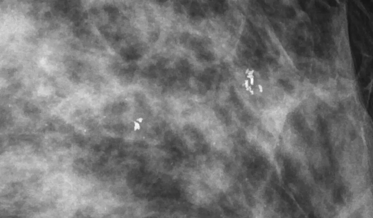
This Agfa white paper examines to what extent the pixel size of full-field digital mammography (FFDM) detectors affects diagnostic image quality and the detection of microcalcifications. To do so, it compares the technical image quality characteristics of several commercial FFDM, retrofit and CR systems with pixel sizes of 50 to 100 µm.
Highlights
- In the 50 to 100 µm range, pixel size alone has no systematic effect on the detectability of small objects.
- Under quantum-noise-limited conditions, SNR is proportional to pixel size; a 76 µm detector may need about 24% more dose than a 100 µm detector to reach the same SNR.
- Pixel size limits the minimum size of detectable detail, but it does not determine the overall resolution of the system.
- DQE is a more comprehensive measure of image quality, covering detector sensitivity, MTF and noise.
- In the CDMAM phantom test, pixel size in the 50 to 100 µm range had no effect on detail contrast visibility.
- MUSICA image processing with the patented Fractional Multiscale Processing (FMP) technology improves diagnostic quality independently of pixel size.
Systems compared
| System | Pixel size (µm) | Pixel matrix | Technology |
|---|---|---|---|
| Agfa DR 24M (on Siemens Nova analog modality) | 76 | 3072 x 3840 | CsI / a-Si TFT |
| Agfa DX-M HM5.0 (on Siemens Nova analog modality) | 50 | 4708 x 5844 | CR Needle IP |
| Fuji Amulet | 50 | 3540 x 4740 | a-Se / optical switch |
| GE Essential | 100 | 2394 x 3062 | CsI / a-Si TFT |
| Hologic Selenia | 70 | 2560 x 3328 | a-Se / TFT |
| Siemens Inspiration | 85 | 2658 x 3318 | a-Se / TFT |
CDMAM test results
| System | kV | A/F | Air Kerma (µGy) | Log(T) 0.1 mm disc | Log(T) 0.25 mm disc |
|---|---|---|---|---|---|
| Agfa DR 24M | 30 | Mo/Rh | 80 | 0.00 | -0.63 |
| Agfa DX-M HM 5.0 | 29 | Mo/Rh | 104 | 0.013 | -0.52 |
| Fuji Amulet | 29 | Mo/Rh | 88 | 0.03 | -0.55 |
| GE Essential | 29 | Rh/Rh | 83 | 0.06 | -0.59 |
| Hologic Selenia | 29 | W/Rh | 100 | 0.08 | -0.62 |
| Siemens Inspiration | 28 | W/Rh | 98 | 0.08 | -0.57 |
A lower value indicates better detectability of low-contrast details.
Paper summary
Early detection of breast cancer often relies on identifying microcalcifications and their geometry, which is why system resolution is considered very important in mammography. FFDM detectors come in different designs: CR with storage phosphor plates, indirect flat panels based on a CsI scintillator, and direct flat panels based on a-Se. CR systems and retrofit panels make it possible to keep using existing mammography modalities.
According to the MTF data, the achievable resolution at high contrast depends mainly on the detector technology: a-Se detectors have the highest MTF at 4 lp/mm, and CR with needle-based plates is nearly on par with CsI detectors. However, the actual visual resolution depends on the ability to display small details with sufficient contrast above the noise.
In addition to the detector, image processing, optimization of the X-ray spectrum through AEC calibration, and the way the user operates the system, including positioning, correct compression, recommended exposure conditions, acceptance tests and regular quality control, play a decisive role in final diagnostic quality.
Source:Agfa(agfahealthcare.com)(opens in a new tab)
Frequently asked questions
Does pixel size of a full-field digital mammography detector in the 50 to 100 µm range affect microcalcification detection?
In the 50 to 100 µm range, pixel size alone has no systematic effect on the detectability of small objects, and the CDMAM phantom test showed no effect on detail contrast visibility in that range.
Why might a 76 µm FFDM detector require more dose than a 100 µm FFDM detector?
Under quantum-noise-limited conditions, SNR is proportional to pixel size; a 76 µm detector may need about 24% more dose than a 100 µm detector to reach the same SNR.
Which metric is more comprehensive than pixel size for evaluating FFDM detector image quality?
DQE is a more comprehensive measure of image quality, covering detector sensitivity, MTF and noise.
Which FFDM detector technology has the highest MTF at 4 lp/mm?
According to the MTF data, a-Se detectors have the highest MTF at 4 lp/mm, and CR with needle-based plates is nearly on par with CsI detectors.
Can MUSICA image processing improve diagnostic quality regardless of FFDM pixel size?
Yes, MUSICA image processing with the patented Fractional Multiscale Processing (FMP) technology improves diagnostic quality independently of pixel size.
Attachments
Comments
No comments yet. Be the first to share your view.
Leave a comment
More to read
Article · Mammography
Agfa Mammography Portfolio
Agfa's vendor-neutral mammography portfolio: Mamoray film, DRYSTAR printers, MUSICA processing, CR Mammo and DR Mammo Retrofit.
Agfa
Article · DR Detectors
Agfa Digital Radiography (DR) Portfolio Overview
Overview of the Agfa DR portfolio: DR 600, DR 800, DX-D 300, DR 400, DX-D 100+, DR 100e, DR Retrofit and DR detectors, all powered by MUSICA image processing.
Agfa

Article · Veterinary
Agfa Equine Digital Radiography with MUSICA 3+ Image Processing
Agfa veterinary DR for equine and large animal imaging with MUSICA 3+ processing, wireless detectors and a low-cost upgrade path from film or CR.
Agfa