

Human Radiology· Hospital
Agfa project
Gelsenkirchen Hospital Case Study: 30% Dose Reduction with DR Retrofit and the DR 14s Detector
EVK Gelsenkirchen moved from Agfa CR to three DR Retrofit systems with DR 14s detectors, gaining faster workflow, consistent images and about 30% dose reduction.
This case study is a reference published by Agfa.
Source: agfahealthcare.com
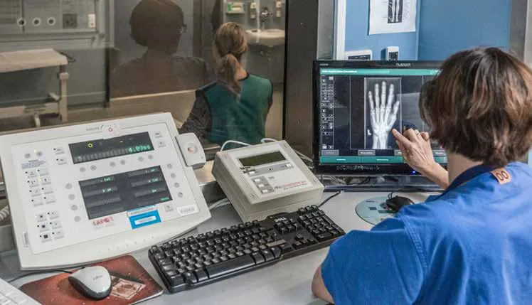
Evangelische Kliniken Gelsenkirchen (EVK), a teaching hospital in Germany affiliated with the University of Duisburg-Essen, moved from Agfa CR plates to three DR Retrofit systems with DR 14s detectors in May 2017. The upgrade resulted in faster work, more consistent image quality and a dose reduction of about 30% without loss of image quality.
Highlights
- Three DR Retrofit systems installed in radiology, the ICU and the outpatient center
- Ultramobile version with a tablet as workstation for the ICU
- Lightweight DR 14s detector, protected against water splashes, with long battery life
- About 30% dose reduction by adjusting exposure parameters and cut-off dose
- Immediate image access, with no need to insert cassettes into a reader
- Significant time savings with a volume of up to 1100 radiographs per week
- MUSICA Chest+ for computational scatter removal in bedside chest radiography
- Smooth installation and effective support from Agfa and distributor Allmedt
Project description
EVK Gelsenkirchen has 433 beds and 15 departments, with about 17,000 admissions and 54,000 outpatient cases per year. The department of diagnostic and interventional radiology and nuclear medicine, with seven radiologists and twelve radiographers, performs more than 20,000 X-ray examinations annually.
The transition from CR plates to DR started in the outpatient center. Agfa and its distributor analyzed the processes and reviewed them with the users, and staff familiarization with the new system went smoothly. In radiology, the radiographer selects the patient from the RIS worklist and the image is available on the monitor immediately after the examination.
In the ICU, patient data and MUSICA are available on a tablet. The image is taken with the mobile X-ray unit and displayed on the tablet via WLAN within seconds, so immediate quality control and retakes are possible without moving the patient again. The system has reduced the radiographers' physical workload and minimized the difficulties of imaging immobile patients.
According to the head of department, DR images are more consistent than CR images and ICU images are easier to assess. The hospital plans to also use the DR 14s in fluoroscopy for free exposures and for bedside chest imaging.
Installed products
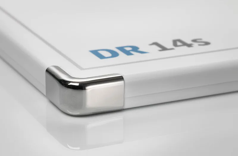 DR Detectors
More projects
View all Human Radiology
Agfa projectHospitalLoma Linda, California, United States
Loma Linda University Medical Center Case Study: Upgrading to DR with the Fast Forward Program
Loma Linda University Medical Center upgraded from CR to DR through Agfa's Fast Forward program with fixed monthly payments and no large upfront capital outlay.
Source: agfahealthcare.com