
Veterinary· Veterinary
Agfa project
Case Study: Livet Equine Veterinary Medical Center, France, with Agfa DR Detectors
France's largest equine hospital made imaging three times faster by moving from CR to Agfa DR 10e C and DR 14e C detectors with the MUSICA workstation.
This case study is a reference published by Agfa.
Source: agfaradiologysolutions.com
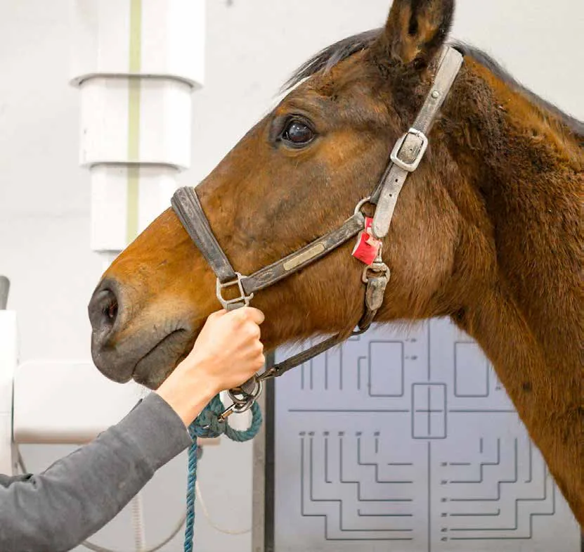
The Livet Equine Veterinary Medical Center in Saint-Michel de Livet, Normandy, is the largest equine hospital in France and performs 50,000 to 60,000 clinical examinations a year. After comparing several suppliers, the center replaced its four CR digitizers with three Agfa DR detectors connected to the MUSICA workstation.
Highlights
- For the same image quality, the radiation dose with the Agfa solution was much lower, and the financial offer was also the best
- With DR, imaging became three times faster; typically 20 to 30 images are taken per horse
- Two DR 10e C detectors (25 x 30 cm) for limbs and field work, and one DR 14e C (35 x 43 cm) in the Bucky for the back, head, neck, shoulder and lungs
- Field work accounts for 40% of the center's consultations
- An Agfa engineer optimized the parameters for each equine anatomical structure to achieve the best quality at the lowest reasonable dose
- Data archived in DICOM format on a NAS
Description
The center has 26 veterinarians and 14 other staff and operates around the clock, mainly treating racehorses and sport horses. It is the only private veterinary clinic in France with CT, MRI and scintigraphy. Its X-ray room is equipped with a column-mounted tube for imaging the horse's entire body and has an adjacent reading room.
According to Dr. Sébastien Caure, a specialist in locomotor disorders, having a monitor in the X-ray room allows images to be checked immediately, and the MUSICA workstation records all of the horse's identification data. Higher quality in complex upper body images has helped to better understand the origin of some conditions.
Solutions used
- DR 10e C detector, 25 x 30 cm
- DR 14e C detector, 35 x 43 cm
- Compact, lightweight detectors with high DQE and optimized pixel size for lower dose
- Excellent connectivity with DICOM-compatible software and imagers
- Long-lasting battery for mobile work
- MUSICA workstation that automatically analyzes each image and applies the best parameters
More projects
View all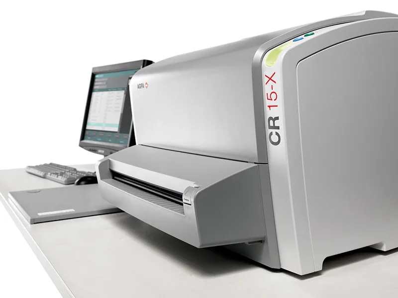 Veterinary
Agfa project2021VeterinaryItaly
Case Study: Clinica della Brughiera, Italy, Equine Imaging with Agfa DR and CR (2021 Edition)
Dr. Alessandro Centinaio's experience with the Agfa DX-D 45C mobile DR and CR 15-X at Clinica della Brughiera and international equestrian events.
Source: agfaradiologysolutions.com
 Veterinary
Agfa project2019VeterinaryItaly
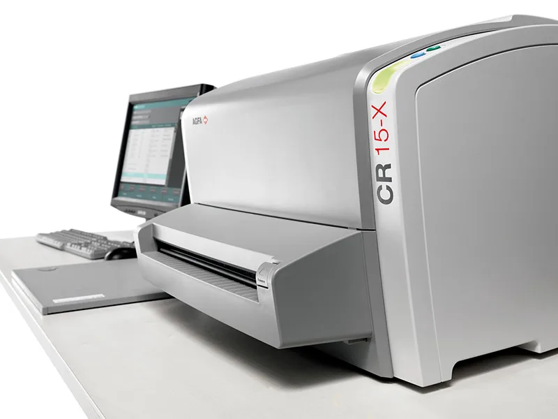
Case Study: Dr. Alessandro Centinaio and Clinica della Brughiera, Italy, with Agfa DR and CR
Equine orthopedic vet Alessandro Centinaio cut pre-purchase imaging time from 1 hour to 20 minutes with the Agfa DX-D 45C mobile DR and CR 15-X.
Source: agfaradiologysolutions.com
 Veterinary
Agfa project2018VeterinaryJyväskylä, Finland
Case Study: AlmaVet Clinic, Finland, with Agfa DR Retrofit
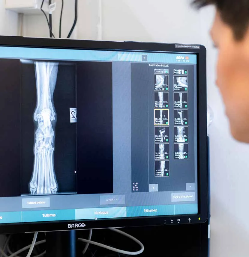
Finnish small animal clinic AlmaVet upgraded from CR to Agfa DR Retrofit with MUSICA processing for more detailed images, lower dose and less invasive procedures.
Source: agfaradiologysolutions.com